

Medical Term For A Freckle
| Lentigo maligna | |
|---|---|
| Other names | Lentiginous melanoma on sun-damaged pare' |
 | |
| Irregular patch well-nigh 10mm square after scrape biopsy which concluded "suspicious of early malignant melanoma". Colour before scrape biopsy was low-cal brown. Mail excision pathology was "Lentigo maligna - Melanoma in situ" | |
| Specialty | Dermatology |
Lentigo maligna is where melanocyte cells have get malignant and grow continuously forth the stratum basale of the peel,[one] but take not invaded below the epidermis.[2] Lentigo maligna is not the same as lentigo maligna melanoma, as detailed beneath. Information technology typically progresses very slowly and can remain in a non-invasive class for years.
Information technology is normally found in the elderly (peak incidence in the 9th decade), on pare areas with high levels of sun exposure similar the face and forearms. Incidence of evolution to lentigo maligna melanoma is depression, about 2.2% to five% in elderly patients.
It is besides known as "Hutchinson'south melanotic freckle".[iii] This is named for Jonathan Hutchinson.[4] [5] The discussion lentiginous comes from the latin for freckle.
Relation to melanoma [edit]
Lentigo maligna is a histopathological variant of melanoma in situ.[vi] Lentigo maligna is sometimes classified equally a very early melanoma,[7] and sometimes as a precursor to melanoma.[8]
When cancerous melanocytes from a lentigo maligna take invaded beneath the epidermis, the condition is termed lentigo maligna melanoma.[2]
Signs and symptoms [edit]
Characteristics include a blue/blackness stain of skin initially. Skin is thin, almost four-5 jail cell layers thick, which is oftentimes related to aging. Histological features include epidermal atrophy and increased number of melanocytes.
Diagnosis [edit]
Get-go dilemma in diagnosis is recognition. As lentigo malignas often nowadays on severely sun-damaged skin, it is oftentimes found amid numerous pigmented lesions – sparse seborrheic keratoses, lentigo senilis, lentigines. It is difficult to distinguish these lesions with the naked eye alone, and even with some difficulty using dermatoscopy. Equally the lentigo maligna is often very big, it often merges with, or encompasses other pare tumors – such equally lentigines, melanocytic nevi, and seborrheic keratosis.
Second dilemma is the biopsy technique. Even though excisional biopsy (removing the entire lesion) is platonic, and advocated by pathologists; practical reason dictates that this should not be washed. These tumors are oft large and presenting on the facial area. Excision of such big tumor would be absolutely contraindicated if the lesion's identity is uncertain. The preferred method of diagnosis is by using a punch biopsy, allowing the physician to sample multiple total thickness pieces of the tumor at multiple sites. While 1 section of the tumor might show benign melanocytic nevus, another section might show features concerning for severe cellular atypia. When cellular atypia is noted, a pathologist might indicate that the entire lesion should be removed. It is at this indicate that one can comfortably remove the unabridged lesion, and thus confirm the terminal diagnosis of lentigo maligna. The size of the dial biopsy can vary from 1 mm to 2 mm, but it is preferable to utilise a punch 1.5 mm or larger. Representative samples of the nearly atypical parts of the nevus should exist biopsied, frequently guided past dermatoscopy.
-

Lentigo Maligna Melanoma, Left Central Malar Cheek marked for biopsy
Handling [edit]
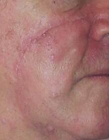
Scar 13 days after excision of coloured patch about 10mm foursquare with 5mm margins from 1cm to correct of base of operations of nose. Length of incision required for skin flap to cover excision site. Scar should lighten and become finer for upward to further half-dozen months if protected from sunday.
The best treatment of lentigo maligna is not clear as it has not been well studied.[ix]
Standard excision is still being washed by about surgeons. Unfortunately, the recurrence charge per unit is high (up to 50%). This is due to the sick-defined visible surgical margin, and the facial location of the lesions (ofttimes forcing the surgeon to use a narrow surgical margin). The use of dermatoscopy can significantly meliorate the surgeon's ability to identify the surgical margin. The narrow surgical margin used (smaller than the standard of care of 5 mm), combined with the limitation of the standard bread loafing technique of fixed tissue histology - result in a high "false negative" mistake rate, and frequent recurrences. Margin controlled (peripheral margins) is necessary to eliminate the simulated negative errors. If breadloafing is utilized, distances from sections should approach 0.i mm to assure that the method approaches complete margin control.
Where the lesion is on the face and either large or 5mm margins are possible, a skin flap or peel graft may be indicated/required. Grafts have their own risks of failure and poor cosmetic outcomes. Flaps tin can require extensive incision resulting in long scars and may be ameliorate done past plastic surgeons (and possibly improve again past those with all-encompassing LM or "suspicious of early cancerous melanoma" experience.
Mohs surgery has been washed with cure rate reported to be 77%.[10] The "double scalpel" peripheral margin controlled excision method approximates the Mohs method in margin control, but requires a pathologist intimately familiar with the complexity of managing the vertical margin on the sparse peripheral sections and staining methods.[eleven]
Some melanocytic nevi, and melanoma-in-situ (lentigo maligna) accept resolved with an experimental treatment, imiquimod (Aldara) topical cream, an immune enhancing amanuensis. In view of the very poor cure rate with standard excision, some surgeons combine the two methods: surgical excision of the lesion, so 3 months handling of the area with imiquimod cream.
Studies seem to disharmonize about the level of certainty associated with using imiquimod.[12] [xiii]
Another treatment to exist considered where standard margins cannot be achieved or cosmetics are a major consideration is ultra-soft ten-ray/grenz-ray radiation.[14]
In the very elderly or those with otherwise express life expectancy, the touch on of major day surgery for excision with 5mm margins and large peel flap could exist worse than doing zero or the possibility of failed treatments with imiquimod or Grenz ray.
References [edit]
- ^ Oakley, Amanda (2011). "Lentigo maligna and lentigo maligna melanoma". DermNet NZ.
- ^ a b Michael Xiong; Ahmad Charifa; Chih Shan J. Chen (2022). "Lentigo Maligna Melanoma". Cancer, Lentigo Maligna Melanoma. StatPearls, National Center for Biotechnology Information. PMID 29489150. Last Update: May 18, 2019.
- ^ Greenish A, Fiddling JH, Weedon D (January 1983). "The diagnosis of Hutchinson's melanotic freckle (lentigo maligna) in Queensland". Pathology. 15 (1): 33–5. doi:x.3109/00313028309061399. PMID 6856341. S2CID 36643652.
- ^ synd/1439 at Who Named It?
- ^ J. Hutchinson. Senile freckle with deep staining - a superficial epithelioma of the cheek. Athenaeum of Surgery, London, 1892, 3: 159.
- ^ McKenna JK, Florell SR, Goldman GD, Bowen GM (Apr 2006). "Lentigo maligna/lentigo maligna melanoma: current state of diagnosis and treatment". Dermatol Surg. 32 (four): 493–504. doi:x.1111/j.1524-4725.2006.32102.ten. PMID 16681656. S2CID 8312676.
- ^ "Precancerous weather of the skin". Canadian Cancer Society . Retrieved 2020-02-26 .
- ^ Fleming, C. (2010). "How to manage patients with lentigo maligna". Melanoma Enquiry. xx: e26. doi:ten.1097/01.cmr.0000382797.99333.66. ISSN 0960-8931.
- ^ Tzellos, T; Kyrgidis, A; Mocellin, S; Chan, A; Pilati, P; Apalla, Z (19 Dec 2014). "Interventions for melanoma in situ, including lentigo maligna". The Cochrane Database of Systematic Reviews. 12 (12): CD010308. doi:10.1002/14651858.CD010308.pub2. PMID 25526608.
- ^ Mikhail, 1000. Mohs Micrographic Surgery. 1991, Saunders, pp. 13-xiv
- ^ Usefulness of the Staged Excision for Lentigo Maligna and Lentigo Maligna Melanoma: The 'Square' Procedure" (J Am Acad Dermatol 1997;37:758-63)
- ^ Li, Lena (2011). "Efficacy of Imiquimod Cream, five%, for Lentigo Maligna After Consummate Excision". Athenaeum of Dermatology. 147 (ten): 1191–5. doi:10.1001/archdermatol.2011.260. PMID 22006136. Retrieved 2 November 2011.
- ^ Powell, A. M. (2009). "Imiquimod and lentigo maligna: a search for prognostic features in a clinicopathological written report with long-term follow-up". British Journal of Dermatology. 160 (5): 994–998. doi:ten.1111/j.1365-2133.2009.09032.x. PMID 19222462. S2CID 25643838.
- ^ Hedblad, Mari-Anne (2012). "Grenz ray treatment of lentigo maligna and early lentigo maligna melanoma". Journal of the American Academy of Dermatology. 67 (i): sixty–68. doi:10.1016/j.jaad.2011.06.029. PMID 22030019.
External links [edit]
-
 Media related to Lentigo maligna at Wikimedia Commons
External links [edit]

Medical Term For A Freckle,
Source: https://en.wikipedia.org/wiki/Lentigo_maligna
Posted by: cobbalkinst.blogspot.com
0 Response to "Medical Term For A Freckle"
Post a Comment